Normal healthy gums are usually described as "coral pink" in color and usually fit to a nice sharp point as they approach where the teeth come together and contact. The healthy gums have a "pebble grain" appearance which is called "stippling". When gums become diseased, they :
| a. | change in color from coral pink to a more reddish color |
| b. | change in form from a nice sharp tapered form to a swollen less tapered form |
| c. | lose their appearance of stippling and become shiny as well as reddened |
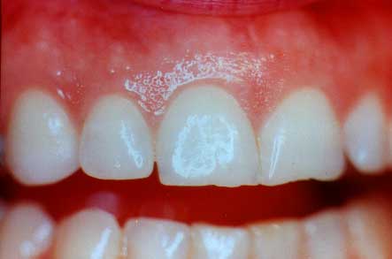
You can see the "coral pink" color, the form where the gums fit in a nice sharp tapered point between the teeth, and where the "stippling" exists.
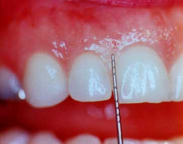
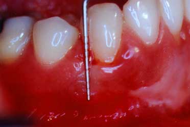
This print shows the probe outside of the gum "sulcus" in order to show just how deep it had gone (2-3 mm.) in this healthy sulcus
Periodontal disease or gum disease looks different in different patients' mouths. However, changes in color form and texture are good visual clues to look for when you are looking for the presence of gum disease.These next photos will show how a deep pocket actually appears.
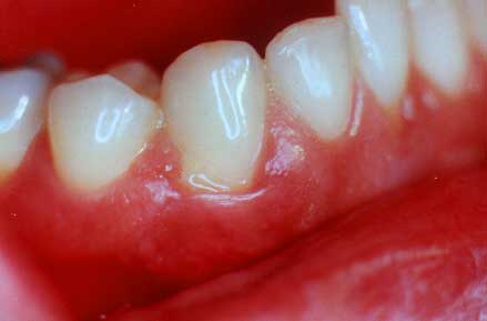
(All diseased areas don't look the same)
The gum tissue has changes in the gum tissue color and form. There is some yellow pus on the gums where the periodontal probe will be inserted in the next photo.
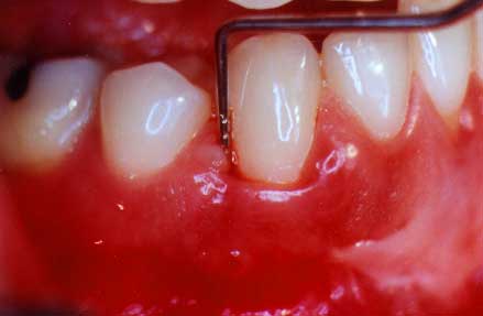
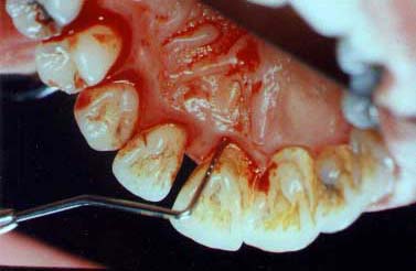
This print shows the probe in the pocket to a depth of 8 millimeters
You can see how deep the probe did actually go into this diseased pocket. This is approximately 8 millimeters and shows how much bone loss has occurred.
Non-surgical therapy is called root planing. Root planing is where the gum tissues are usually anesthetized with a novocaine anesthetic and the dentist or hygienist will scrape the diseased tooth root until it is clean and no longer contains bacterial waste products. Root planing may sometimes be enhanced with antibiotics that are taken orally or placed into the gum "pockets".
Not all cases of gum disease will have antibiotic enhancements as a part of their root planing therapy. After root planing, the gum tissues are usually tender or sore, but not usually painful. When the periodontist determines the gum disease aggressiveness, the periodontist can determine the modifications in the root planing treatments that best fit your case.
(pre-treatment - roof of the mouth view)
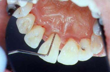
Here you can see the silver periodontal probe which goes 5 mm. into the pocket. There is much bleeding and infection present.
The gum tissues are pink in color and there has been some tissue shrinkage. The gums are healthy with a probing depth of 3 mm.
Gum surgery is done to eliminate diseased pockets which still exist after all healing has been achieved following non-surgical root planing. There are however a small number of cases which will not benefit from non-surgical root planing and surgery will be recommended without having root planing done. Surgery is most frequently done with a novocaine local anesthetic. A periodontal dressing may be placed after the surgery in order to protect the tissues. There may be some pain after periodontal surgery.
When surgery is done and diseased tissue is removed, the teeth will usually have more root surface exposed. This amount of exposure represents the amount of root surface which lost its gum and bone support from the gum disease. The surgery is not causing unnecessary damage.
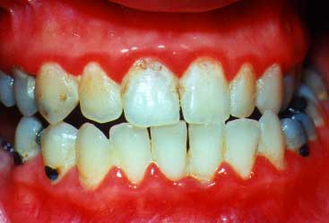
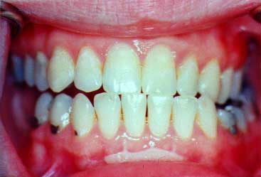
(lips are retracted)
This view shows very inflamed with changes in color, form, and the presence of recession between the teeth. Gum surgery will be needed because root planing alone cannot make her gums healthy.
The gums are now healthy. The pink color and restored contours are evidence of that health. She can now maintain her teeth for all of her life.